Annexin V-FITC staining to detect cell apoptosis
Licia Miller Product Manager
Procedure for early detection of apoptosis using Annexin V-FITC staining and optional propidium iodide ( PI ) .
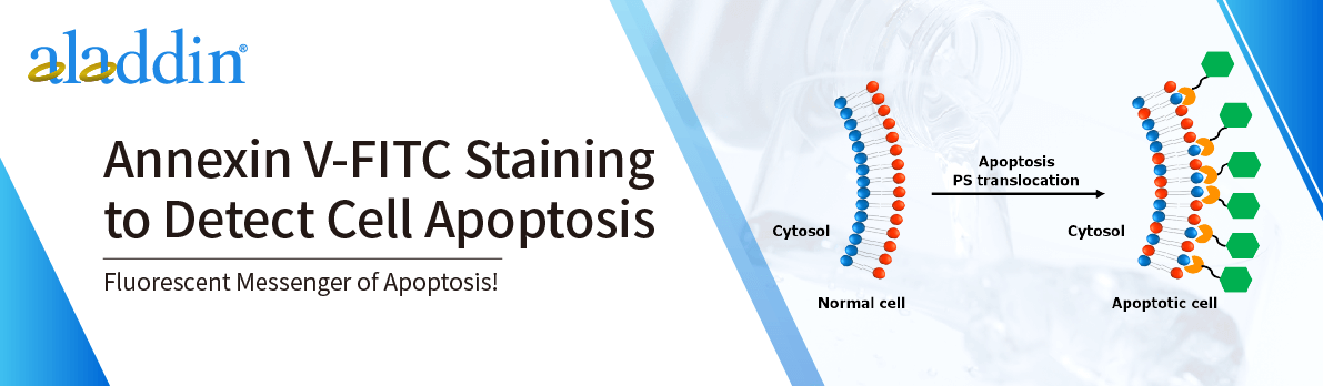
of phosphatidylserine ( PS ) residues (normally hidden in the plasma membrane) to the cell surface is an early event in apoptosis and can be used to detect and measure apoptosis. During apoptosis, PS is transferred from the cytoplasmic face of the plasma membrane to the cell surface. Annexin V has a strong Ca2+-dependent affinity for PS and can therefore be used as a probe for detecting apoptosis.
This is an example protocol for a PS exposure assay using Annexin V, based on the protocol provided in the Annexin V-FITC Apoptosis Detection Kit. Please note that when using a specific kit, you should always use the protocol provided on the datasheet, as it is designed for optimal performance with the product.
Reagents:
- 1× Annexin V Binding Buffer
-Optional : Propidium Iodide
- 2% Formaldehyde
-Optional : Trypsin (for adherent cells)
Phase 1 Incubate cells with Annexin V-FITC
- Cell incubation (general purpose)
Experimental steps
1. Induce cell apoptosis by a desired method.
2. Collect cells by centrifugation.
• You will need to collect 1-5×10⁵ cells, depending on the volume required for your experiment.
3. Resuspend the cells in 500 µL 1 × Annexin V Binding Buffer.
4. Add 5 µL of Annexin V-FITC.
• If you want to analyze PI staining later, you can also add 5 µL of propidium iodide ( PI ) at this step.
5. Incubate at room temperature in the dark for 5 minutes.
- Adherent cells
Experimental steps
1. Induce cell apoptosis by a desired method.
2. Collect cells by centrifugation.
• You will need to collect 1-5×10⁵ cells, depending on the volume required for your experiment.
3. Gently digest the cells with trypsin and wash the cells once with serum-containing medium.
• Note: This should be done prior to incubation with Annexin V-FITC (steps 4 and 5).
4. Resuspend the cells in 500 µL 1 × Annexin V Binding Buffer.
5. Add 5 µL of Annexin V- FITC.
• If you want to analyze PI staining later, you can also add 5 µL of propidium iodide ( PI ) at this step.
6. Incubate at room temperature in the dark for 5 minutes.
Phase 2 Analysis of Annexin V-FITC binding
Annexin V-FITC binding was analyzed by flow cytometry.
Experimental steps
1. Analysis of Annexin V-FITC binding by flow cytometry.
• Ex = 488 nm and Em = 350 nm using FITC signal detector (usually FL1).
2.If propidium iodide was added, analyze PI staining using a phycoerythrin emission signal detector (usually FL2).
For more product details, please visit Aladdin Scientific website.