My Cart
You have no items in your shopping cart.
Creating an account has many benefits:
Licia Miller Product Manager
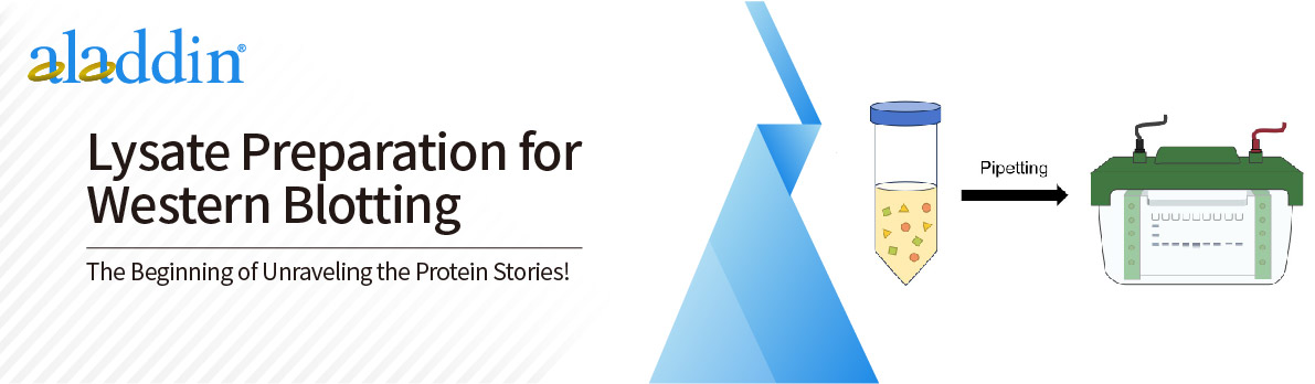
This method provides a recipe of lysate and buffer preparation for lysing cells or tissues for western blot analysis.
The buffer solution formula is as follows:
( 1 ) 1×1% SDS pyrolysis buffer
- 10 1 mM Tris-HCl (pH 8.0)
- 1% SDS
- 1 mM Sodium orthovanadate
- ddH2O
( 2 ) 2× sample buffer
- 62.5 1 mM Tris-HCl (pH 6.8)
- 2% SDS
- 0.01% bromophenol blue
- 25% Glycerin
- 710 mM β-mercaptoethanol
- ddH2O
• Cell samples
Ⅰ. Preparation of 1% SDS thermal lysis buffer (denaturation)
Experimental Steps
1. Discard the culture medium in the culture flask and wash once with pre-cooled PBS.
2. Add 3 ml of pre-cooled PBS to each culture flask and collect the cells using a cell scraper.
3. Add 12 ml of pre-chilled PBS to ensure that all cells are detached from the culture flask.
4. Transfer the collected cells to a 50 ml centrifuge tube and centrifuge at 300 ×g for 5 minutes.
5. Discard the supernatant and wash twice with pre-cooled PBS.
6. Heat 1% SDS pyrolysis until foaming.
7. Use a pipette to add 1% SDS hot cell lysis buffer, resuspend the cells , and let stand for 10 to 20 minutes.
8. Use an ultrasonic cell disruptor to break up all cell clumps until the lysate becomes clear. The ultrasonic time is usually 3 s, interval 10 s, ultrasound 5-15 times, ultrasound power: 40 kW.
9. Centrifuge at 15,000-17,000 ×g for 5-10 minutes and discard the cell pellet.
Experimental Steps
1. Discard the culture medium in the culture flask and wash once with pre-cooled PBS.
2. Add 3 ml of pre-cooled PBS to each culture flask and collect the cells using a cell scraper.
3. Add 12 ml of pre-chilled PBS to ensure that all cells are detached from the culture flask.
4. Transfer the collected cells to a 50 ml centrifuge tube and centrifuge at 300 ×g for 5-10 minutes.
5. Discard the supernatant and wash twice with pre-cooled PBS.
6. Add RIPA Buffer according to the cell volume and resuspend the cells . Place on ice for 15 minutes.
7. Use an ultrasonic cell disruptor to break up all cell clumps until the lysate becomes clear. The recommended ultrasonic time is 3 s, interval 10 s, ultrasound 5-15 times, ultrasound power: 40 kW.
8. Centrifuge at 15,000-17,000 ×g for 5-10 minutes and discard the cell pellet.
• Tissue samples
Ⅰ. Preparation of 1% SDS thermal lysis buffer ( denaturation )
Experimental Steps
1. Use pre-cooled scissors to cut the frozen tissue into pieces. Then use a pre-cooled mortar to grind the tissue into powder.
2. Heat 1% SDS pyrolysis until foaming.
3. Use a pipette to add 1% SDS hot cell lysis buffer and resuspend the cells. Leave for 10 to 20 minutes.
4. Use an ultrasonic cell disruptor to break up all cell clumps until the lysate becomes clear. The general ultrasonic time is 3 s, interval 10 s, ultrasound 5-15 times, ultrasound power: 40 kW.
5. Centrifuge at 15,000-17,000 ×g for 5-10 minutes and discard the cell pellet.
II. RIPA Lysate Preparation
Experimental Steps
1. Use pre-cooled scissors to cut the frozen tissue into pieces. Then use a tissue grinder to grind the tissue into powder.
2. Add RIPA buffer according to the amount of tissue and resuspend the tissue. Place on ice for 15 minutes.
3. Use an ultrasonic cell disruptor to break up all cell clumps until the lysate becomes clear. Generally , the ultrasonic time is 3s, with an interval of 10 s, ultrasound 5-15 times, ultrasound power: 40 kW.
4. Centrifuge at 300 × g for 5-10 minutes and discard the tissue pellet.
For more product details, please visit Aladdin Scientific website.