My Cart
You have no items in your shopping cart.
Creating an account has many benefits:
Summary
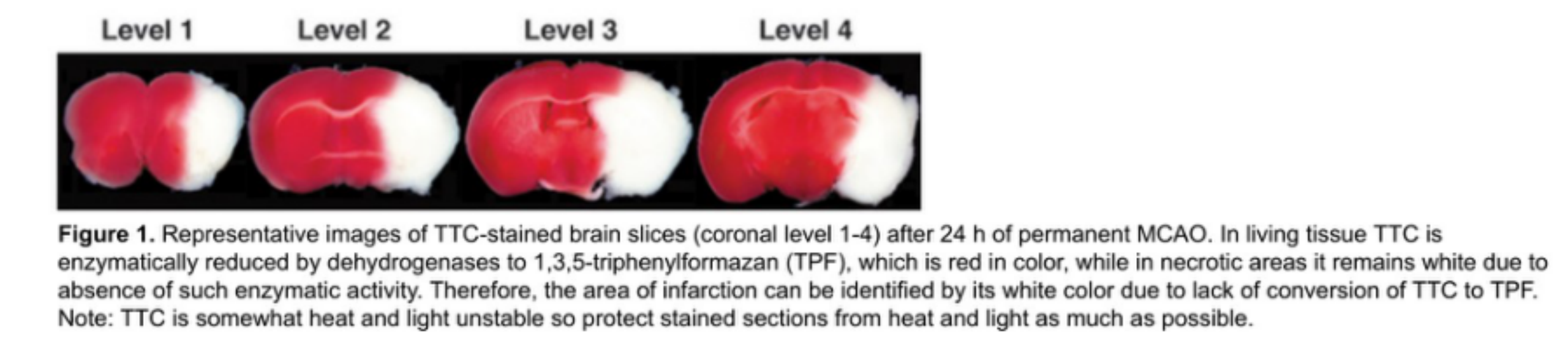
TTC (2,3,5-triphenyltetrazolium chloride) is a fat-soluble photosensitive complex. It is a proton receptor for the pyridine-nucleoside structural enzyme system in the respiratory chain and reacts with dehydrogenase in normal tissues to give a red color, whereas dehydrogenase activity is decreased in ischemic tissues, which cannot react, and therefore does not produce a change to a pale color. Therefore, TTC staining is used to stain mammalian tissues to detect ischemic infarction.
Operation method
TTC staining test (TTC)
Principle
TTC (2,3,5-triphenyltetrazolium chloride) is a fat-soluble photosensitive complex. It is a proton receptor for the pyridine-nucleoside structural enzyme system in the respiratory chain and reacts with dehydrogenase in normal tissues to give a red color, whereas dehydrogenase activity is decreased in ischemic tissues and cannot react, so it does not produce a change to a pale color.
Materials and Instruments
2,3,5-triphenyltetrazolium chloride solution; coronal sectioned brain slots; blades; sodium pentobarbital; syringes; and surgical instruments for brain extraction.
Move
1. Anesthetize the mice, break the neck and take out the brain, put the brain into the brain tank. Put the brain into -80 ℃ refrigerator for 1~2 min or -20 ℃ refrigerator for 20 min, and freeze the brain a little bit hard to facilitate the sectioning. 2.
2. Coronal sectioning of the brain was performed in four 2 mm slices, taking care not to damage the integrity of the brain slices. The first slice was made at the midpoint of the line between the anterior cerebral level and the optic intersection; the second slice was made at the optic intersection; the third slice was made at the funicular peduncle; and the fourth slice was made between the funicular peduncle and the caudal level of the posterior lobe. 3.
3. Put the brain slices into 2% TTC solution (dissolved in PBS), and put them into a 37 ℃ constant temperature box for 15 min~30 min, and turn the slices halfway to make the staining uniform.
4. Take photographs for observation, or put the stained brain slices into 4% formalin solution and fix and store them for subsequent quantitative photographic detection of infarcted areas.
Caveat
1. TTC staining should be kept away from light.
2. Animal sampling does not require perfusion sampling, and the brain can be taken out directly. The brain tissue is soft, so care should be taken to ensure the integrity of the brain.
3. When making brain slices, it is better to freeze the brain beforehand, and it is not easy to be damaged.
4. Too dark or too light staining may be related to the staining time, which can be adjusted appropriately.
Common Problems
1. What should I do if the brain is easily damaged when slicing the brain?
A: Make sure there is no water in the brain tank, put the brain into the brain tank quickly and put it into the -80 ℃ refrigerator for 1~2 min or -20 ℃ refrigerator for 20 min, when removing the brain slices, it is easier to remove the brain slices from the brain tank by lifting the two blades out together.
2. Why is there no white infarct area in TTC staining?
A: Consider whether the modeling is successful, whether to stain at 37 ℃ for 15 min, and the whole staining process must be protected from light.
For more product details, please visit Aladdin Scientific website.