My Cart
You have no items in your shopping cart.
Creating an account has many benefits:
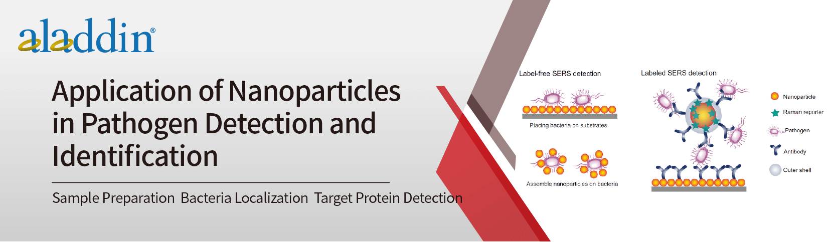
Rapid pathogen detection and identification are of considerable importance in the early stages of food safety and treatment to avoid drug resistance. Modern laboratories use a number of molecular biology assays to distinguish these drug-resistant pathogenic strains from foodborne pathogens, including whole genome sequencing, modern PCR, multiplex PCR, quantitative PCR (qPCR), and droplet PCR. While real-time PCR (RT-PCR) and qPCR can be used to continuously monitor changes in bacteria, they are too expensive to be applied in resource-limited settings, whereas qualitative assays are more cost-effective and dominate the field. Therefore, advanced and novel pathogen detection and identification techniques have attracted much attention. In this regard, nanoparticles (NPs) have come into people's field of vision because they are more sensitive, accurate, and reliable in qualitative detection. There are many research and application reports on the use of nanoparticles for pathogen detection and identification.
Sample Preparation
In the past, if we used gold-standard technology to test critically ill patients who needed specific antibiotics within 24 hours, it could have taken a long time. However, with the help of a fast and highly simple extraction process called immunomagnetic separation, the time to concentrate microorganisms from various clinical samples can be shortened to a few hours. Magnetic nanoparticles (MNPs) have been widely used in sample preparation due to their excellent magnetic properties, low cost, analytical versatility, and high capture efficiency. For example, Kearns et al. used lectin-functionalized silver-coated MNPs and optically active antibody-coated silver-coated NPs to isolate and detect three bacterial pathogens (including methicillin-resistant Staphylococcus aureus, MRSA). Furthermore, Cowger et al. developed a novel drug susceptibility test utilizing NPs - mediated mass spectrometry that takes less than 50 min and can differentiate wild-type from drug-resistant strains (Fig. 1).

Figure 1: Schematic illustration of bacterial isolation using protein-adsorbed magnetic nanoparticles
MRI Localization of Bacteria
Considering bacterial resistance and organ damage, the application of MRI as noninvasive imaging has great potential to localize bacteria early in infection. This technique has been used to visualize S. aureus infection in endocarditis, osteomyelitis, and soft tissue infection models. However, these models examine inflammation, not the causative agent of infection. There has been one study using iron particles to directly visualize S. aureus, but the lack of specificity has limited the application of this method. The development of specific molecular imaging probes is underway, including using iron NPs coupled to IgG to visualize intracellular pathogens. Scientists have reported that extrapulmonary mycobacterial infection is specifically detected by particles coated with antibodies to the surface of M. tuberculosis.
Detection of the target protein by IPCR
In the detection of antigens and antibodies that cannot be amplified in number or sequence, a highly sensitive technique, immuno-PCR (immunopcr, IPCR), was introduced into these studies, which combines immunology and PCR to detect proteins of interest. Typically, antibodies are conjugated to a known DNA sequence, then used to target the target antigen in the sample (Figure 2), and PCR is finally used to amplify the sequence. With the use of IPCR, the detection limit of typical ELISA can be increased to 100-10000 times. IPCR has expanded from its use in research to diagnostic laboratories for the detection of proteins with greater sensitivity. Many studies have demonstrated that gold NPs can enhance the sensitivity of IPCR. Antibodies bind more easily to gold NPs than to DNA directly. Moreover, gold NPs
Can be combined with more DNA molecules, thus further improving the sensitivity of IPCR. Using a similar strategy, Chen and colleagues combined IPCR with biologically barcoded amplification using gold NPs to detect Hantaan virus nucleocapsid proteins.

Figure 2: IPCR for detection of target antigens. Where a DNA-conjugated antibody is used to detect the antigen, PCR amplification of the DNA sequence can quantitatively determine the presence of the antigen
Applications in the Food Industry
Foodborne illness has been a serious threat to public health for decades and remains a major public health challenge. Nanoparticle-based biosensors have been developed to detect pathogens, including the use of magnetic NPs, silver NPs, and silver nanoshells. However, these methods lack the ability to detect multiple organisms in a single assay. Recently, three dye-doped fluorescent silica NPs and quantum dots (QDs) were reported for the detection of various bacteria. But there are still downsides to this strategy. Fluorescent detection methods require the use of fluorescent probes, which are susceptible to photobleaching and careless handling, while quantum dots are difficult and expensive to manufacture and complex to modify on the surface.
References
1. Van Giau, V., An, SSA, & Hulme, J. (2019). Recent advances in the treatment of pathogenic infections using antibiotics and nano-drug delivery vehicles. Drug design, development, and therapy, 13, 327. https ://www.tandfonline.com/doi/full/10.2147/DDDT.S190577
2. Syed, MA, & Bokhari, S. (2011). Gold nanoparticle-based microbial detection and identification. Journal of biomedical nanotechnology, 7(2), 229-237. https://doi.org/10.1166/jbn.2011.1281
3. Wang, C., & Irudayaraj, J. (2008). Gold nanorod probes for the detection of multiple pathogens. Small, 4(12), 2204-2208. https://doi.org/10.1002/smll.200800309
4. Chen, L., Wei, H., Guo, Y., Cui, Z., Zhang, Z., & Zhang, XE (2009). Gold nanoparticle enhanced immuno-PCR for ultrasensitive detection of Hantaan virus nucleocapsid protein. Journal of immunological methods, 346(1-2), 64-70. https://doi.org/10.1016/j.jim.2009.05.007
5. Cowger, TA, Yang, Y., Rink, DE, Todd, T., Chen, H., Shen, Y., ... & Xie , J. (2017). Protein-adsorbed magnetic-nanoparticle-mediated assay for rapid detection of bacterial antibiotic resistance. Bioconjugate Chemistry, 28(4), 890-896. https://doi.org/10.1021/acs.bioconjchem.7b00016
6. Kearns, H., Goodacre, R., Jamieson, LE, Graham, D., & Faulds, K. (2017). SERS detection of multiple antimicrobial-resistant pathogens using nanosensors. Analytical chemistry, 89(23), 12666 -12673. https://doi.org/10.1021/acs.analchem.7b02653
7. Liu, J., Bai, R., Li, Y., Staedtke , V., Zhang, S., van Zijl , PC, & Liu, G. (2018). MRI detection of bacterial brain abscesses and monitoring of Antibiotic treatment using access. Magnetic resonance in medicine, 80(2), 662-671. https://doi.org/10.1002/mrm.27180
8. Hoerr , V., Tuchscherr , L., Hüve , J., Nippe , N., Loser, K., Glyvuk , N., ... & Klingauf , J. (2013). Bacteria tracking by in vivo magnetic resonance imaging. BMC biology, 11(1), 63. https://bmcbiol.biomedcentral.com/articles/10.1186/1741-7007-11-63
9. Moro, L., Turemis , M., Marini, B., Ippodrino , R., & Giardi , MT (2017). Better together: Strategies based on magnetic particles and quantum dots for improved biosensing. Biotechnology advances, 35( 1), 51-63. https://doi.org/10.1016/j.biotechadv.2016.11.007