Dye-Aggregation

The absorption of light results in dye molecules entering an electronically excited state, which can store energy only for a brief period and can be radiated again during fluorescence after the excited state's lifetime.
When dye molecules are in a solution, they are referred to as point dipoles or oscillators. They do not influence each other if their distances are large enough.
At an average distance of approximately 5-10 nm between the chromophores, the "radiation field" of the oscillators only has an impact. Förster's resonance energy transfer (FRET) model describes this type of interaction between two dye molecules.
When the distance between the chromophores decreases even further, as in a highly concentrated solution, the individual oscillators can significantly affect each other through electrostatic interaction. The absorption and fluorescence behavior of a dye solution can be strongly affected by the intermolecular interaction among its individual dye molecules.
Rhodamine 6G in water
The concentrated aqueous solution of rhodamine 6G exhibits a shoulder at the short-wave flank of the primary absorption band in its UV/Vis spectrum. Upon diluting the solution by altering the concentration (c) and increasing the optical path length (d) of the cuvette, the same absorbance would be expected, according to the Lambert-Beer law. The following spectra can be observed:

The presence of an isosbestic point, where the alteration in concentration of all the substances involved is consistent, dE/dc = 0, indicates that two or more species are in a dynamic equilibrium with each other.
![]()
The dissociation or association/complexation constant can be determined experimentally. Conduct a dilution series in which the solution's dilution is always compensated by the path length change. Subsequently measure the absorbance at the (monomer) maximum, and use the dilution factor and the initial sample concentration to calculate the "effective extinction coefficient". The concentration of this sample is determined by analyzing the UV spectrum of a highly diluted solution where dimerization does not occur. Due to the additive behavior of absorption of different species in the Lambert-Beer law, it is possible to ascertain an effective extinction or extinction coefficient by leveraging knowledge of the underlying reaction. By varying the dissociation constant and graphically analyzing the results, a straight line can be obtained. This line provides information on the extinction coefficients of both the monomer and dimer.
Hydrophobic interaction
Organic dyes tend to aggregate in high ionic strength solvents or water due to intermolecular van der Waals forces, also known as the "hydrophobic interaction". Lipophilic dye molecules reduce their hydrate shell's surface area by avoiding hydrophilic water molecules. This phenomenon is also the reason for dyes adsorbing onto glass surfaces or non-specifically bonding to substrate molecules.
The possibility of forming dimers or higher aggregates depends on:
The dye concentration: greater concentration leads to higher aggregation.
The solvent: in water or methanol, rather than ethanol or other organic solvents exhibits elevated occurrences of aggregation. The absorption spectra of ATTO 565, containing equal concentrated solutions in aqueous PBS buffer (pH 7.4) and ethanol with trifluoroacetic acid (TFAc), were compared and impressively demonstrated this phenomenon.

In organic solvents like chloroform, the presence of electrolytes (salts) may cause ion pairs (dye cation and counter ions) to form, which
Temperture: Higher temperatures can hinder aggregation due to increased thermal movement.
The molecular structure of the dye: Dyes with hydrophilic groups (e.g. ATTO 488, ATTO 532, ATTO 542, etc.) typically do not aggregate in aqueous solution, while more hydrophobic dyes like ATTO Rho6G, ATTO Rho11, and ATTO Rho12 do.
As this is a dynamic equilibrium, the dimers can be reconverted to monomers by diluting the solution. The "monomer spectrum" is reached when the measured absorption spectrum does not change with further dilution and corresponding increase in the optical path length. For most hydrophobic ATTO dyes, this is the case at an absorbance of approximately 0.04.
Intramolecular interaction in protein conjugates / DOL determination
The NHS ester dye reaction with a protein's amino groups can result in the formation of closely adjacent dye conjugates that interact with each other. This interaction can cause a significant alteration in the absorption spectrum. This effect is evident in the case of the ATTO 565 streptavidin conjugate.
An additional short-wave absorption band is observed in the conjugate spectrum, similar to the "dimer band" of an aqueous dye solution with a high concentration. This is due to the intramolecular interaction of the covalently bound dye molecules, causing no change in the absorption spectrum of the conjugate solution when diluted.
The "Procedures" section of our user guide and protocols describes how to determine the degree of labeling (DOL) in such cases.
There are two main types of aggregates:
H aggregates (H = hypsochrome)
Aggregation of this nature arises when two or more dye molecules are aligned parallel to each other with their transition dipole moments (typically along the longitudinal axis of the chromophoric system in the S0-S1 transition). Unlike monomer absorption, this mode of aggregation results in a hypsochromically shifted absorption band.
The spatial proximity between the two molecules results in electronic orbitals exerting an influence on each other. Consequently, they must be treated as a single unit. The quantum mechanical rules allow absorption at shorter wavelengths due to a split in energy levels leading to higher energy levels. Fluorescence is no longer viable since the fast internal conversion (IC) occurs from this higher lying excitation state.
J aggregates (according to E.E. Jelley)
This type of aggregation leads to a shift to longer wavelengths of the absorption band, which results in a significant decrease in the band's half-width.
J aggregates are frequently found in polymethine dyes, including cyanines, merocyanines or similar chromophores. Jelley and Scheibe independently observed this phenomenon for the first time on the dye pseudoisocyanine.
Various methods have been suggested to model the "supramolecular polymer" created by the union of individual dyes. The easiest approach to describe the molecular relationships is that each molecule aligns with one another, resulting in in-line transition dipole moments. As a result of the collective consideration of the molecules, the energy levels split. The allowed quantum-mechanical transition has now decreased in energy, and this accounts for the shift towards longer wavelengths in the absorption band.
The aggregation can be greatly impacted by the solvent composition, addition of salts or additives, and dye concentration. In optimal conditions, the narrow absorption band is visible in the UV/Vis spectrum. Unlike H aggregates, this type of aggregation can exhibit fluorescence, especially at lower temperatures. Nevertheless, the maximum emission band is only slightly red-shifted compared to the absorption maximum.
Depending on experimental conditions, literature reports a "broadening" of the absorption band, explained by the inclusion of the forbidden electron transfer of the J aggregate.
ATTO 488 marked phospholipids
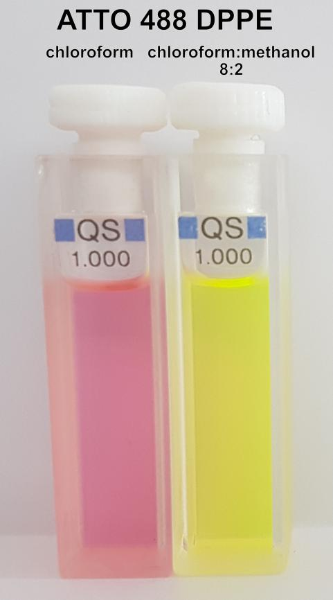
Solutions of ATTO 488 labelled phospholipids in chloroform are initially surprising due to their unexpected colour. Instead of the expected light yellow with bright green fluorescence, the solution appears pink to magenta, likely due to J aggregates. Diluting the solution with methanol causes the colour to change to yellow and become strongly fluorescent. Altering the solvent composition reduces the aggregates.
The figures illustrate the solutions of DPPE, marked with ATTO 488, in pure chloroform and a solvent mixture of chloroform/methanol (8:2, V/V).
Both solutions are displayed in normal daylight on the left, while the green fluorescence of the solution mixed with methanol is clearly visible on the right when exposed to UV light (366 nm).
Reference
E. Jelley, Spectral Absorption and Fluorescence of Dyes in the Molecular State, Nature 138, 1009 (1936).
2. G. Scheibe, Über die Veränderlichkeit der Absorptionsspektren in Lösungen und die Nebenvalenzen als ihre Ursache, Angewandte Chemie 50, 212 (1937).
3. T. Förster, Energiewanderung und Fluoreszenz, Die Naturwissenschaften 33, 166 (1946).
4. M. Kasha, H.R. Rawls, M. Ashraf El-Bayoumi, The Exciton Model in Molecular Spectroscopy, Pure Appl. Chem. 11, 371 (1965).
5. O. Valdes-Aguilera, D.C. Neckers, Aggregation Phenomena in Xanthene Dyes, Acc. Chem. Res. 22, 171 (1989).
6. J. Hernando et al., Excitonic Behavior of Rhodamine Dimers: A Single-Molecule Study, J. Phys. Chem. A 107, 43 (2003).
7. V.I. Gavrilenko, M.A. Noginov, Ab initio study of optical properties of rhodamine 6G molecular dimers, J. Chem. Phys. 124, 0044301 (2006).


