Gold Nanoparticles: Properties and Applications

Sandra Forbes
Product Manager
Introduction
For centuries, scientists have employed colloidal gold nanoparticles, exploiting their captivating hues that stem from their intricate interactions with visible light. In recent times, the extraordinary optoelectronic characteristics of these nanoparticles have sparked intensive research and found their way into cutting-edge technologies, encompassing diverse fields like organic photovoltaics, sensory probes, therapeutic modalities, drug delivery systems for biological and medical applications, electronic conduction materials, and catalysis. By adjusting factors such as their size, shape, surface chemistry, or aggregation status, the optical and electronic properties of gold nanoparticles can be finely tuned to suit specific purposes.
Optical & Electronic Properties of Gold Nanoparticles
The behavior of gold nanoparticles in response to light is heavily influenced by their surrounding environment, size, and geometric configuration. When a light beam's oscillating electric fields approach a colloidal gold nanoparticle, they engage with the free electrons, initiating a synchronized oscillation of electronic charge that resonates with the frequency of visible light. This resonant oscillation phenomenon is termed surface plasmon resonance. In the case of small (~30 nm), uniformly sized gold nanoparticles, this resonance triggers the absorption of blue-green light (~450 nm), while reflecting red light (~700 nm), resulting in a vibrant red hue. As particle sizes escalate, the absorption related to surface plasmon resonance shifts towards longer, redder wavelengths, causing red light to be absorbed and blue light reflected, producing pale blue or purple hues (Figure 1). As particles grow closer to their bulk size, the resonance wavelengths migrate into the infrared spectrum, reflecting most visible wavelengths and imparting a clear or translucent appearance to the nanoparticles. By manipulating the size or shape of these nanoparticles, the surface plasmon resonance can be tailored, yielding particles with customized optical properties suited for diverse applications.

Figure 1. Colors of various sized monodispersed gold nanoparticles
A similar transformation occurs when an excess of salt is introduced into a gold nanoparticle solution. This addition neutralizes the surface charge of the nanoparticles, prompting them to coalesce. Consequently, the solution's color shifts from red to blue. To mitigate aggregation, the versatile surface chemistry of gold nanoparticles facilitates their coating with polymers, small molecules, and biological recognition elements. This surface modification strategy expands the applicability of gold nanoparticles across chemical, biological, engineering, and medical domains. Table 1 outlines some of the characteristic properties of gold nanoparticles.

Table 1. Product Properties of Gold Nanoparticles
Applications
The applications of gold nanoparticles are rapidly expanding, encompassing diverse fields such as:
1. Electronics: Gold nanoparticles are indispensable in electronics, serving as conductors in everything from printable inks to microchips. As electronics continue to shrink, nanoparticles play a crucial role in chip architecture, connecting resistors, conductors, and other components.
2. Photodynamic Therapy: Near-infrared (NIR) absorbing gold nanoparticles, including nanoshells and nanorods, harness light energy at wavelengths between 700 and 800 nm to generate heat, enabling targeted tumor ablation. This hyperthermia therapy leverages the rapid heating of nanoparticles upon light exposure to destroy tumor cells.
3. Therapeutic Agent Delivery: Gold nanoparticles' high surface-to-volume ratio allows for the attachment of numerous molecules, including therapeutic agents, targeting moieties, and anti-fouling polymers, making them ideal for drug delivery.
4. Sensors: Gold nanoparticles find use in diverse sensing applications. Colorimetric sensors utilizing gold nanoparticles can assess food safety, while surface-enhanced Raman spectroscopy employs gold nanoparticles as substrates to detect vibrational properties of chemical bonds, enabling label-free detection of proteins, pollutants, and other molecules.
5. Probes: Gold nanoparticles exhibit unique light-scattering properties, producing vibrant colors under dark-field microscopy, making them invaluable for biological imaging. Their high density also renders them suitable as probes in transmission electron microscopy.
6. Diagnostics: Gold nanoparticles facilitate biomarker detection in various medical conditions, including heart diseases, cancers, and infections. They are also prevalent in lateral flow immunoassays, a technology familiar to households through home pregnancy tests.
7. Catalysis: Gold nanoparticles serve as catalysts in numerous chemical reactions, enhancing selective oxidation processes and, in some cases, facilitating reduction reactions like nitrogen oxide reduction. Research is ongoing to harness their catalytic properties in fuel cells, with potential applications in automotive and display industries.
Aladdin Quality Advantage
We are delighted to present an extensive range of gold nanoparticles, tailored exclusively for cutting-edge applications across life science and materials science domains. Our offering encompasses gold nanoparticles with diameters spanning from 5 nm to 400 nm, featuring diverse surface functionalities and dissolved in a multitude of solvents.
Departing from the conventional use of strong reducing agents like sodium citrate or sodium borohydride for spherical gold nanoparticle synthesis, Aladdin boasts a proprietary process and formulation that yields highly spherical nanoparticles without resorting to such harsh chemicals. These nanoparticles stand out from their peers, offering numerous advantages, among which are:
1. A narrow size distribution, rigorously verified through Dynamic Light Scattering (DLS) and Transmission Electron Microscopy (TEM) analyses. Each batch undergoes stringent quality control, including DLS and UV-Vis spectroscopy evaluations (as illustrated in Figure 2), ensuring consistency and precision.

Figure 2. DLS & UV-Vis spectra showing precise gold nanoparticles from Aladdin.
2. Consistent Size and Shape — <10% CV (coefficient of variance) even above 100 nm. Example of 5 nm and 400 nm nanoparticles are shown below in Figure 3.
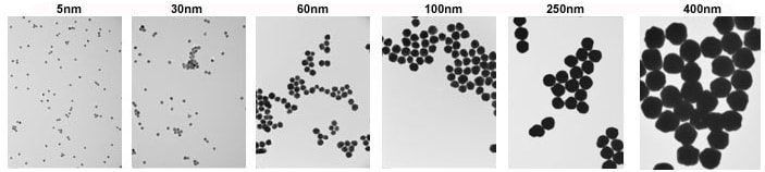
Figure 3. TEM images of 5 nm (left) and 400 nm (right) gold nanoparticles with <8% CV.
Gold NanoUrchins
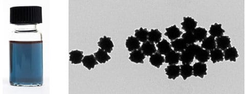
Figure 4. TEM of 100 nm Gold NanoUrchins
Gold NanoUrchins exhibit distinctive optical characteristics when juxtaposed with spherical gold nanoparticles of identical core diameters. Their spiky, irregular surfaces induce a red shift in the surface plasmon resonance (SPR) peak and generate a more pronounced enhancement of the electromagnetic field at the tips of their protrusions, surpassing that observed in spherical counterparts. For instance, 100nm spherical gold nanoparticles display an SPR peak at 570nm, whereas 100nm Gold NanoUrchins exhibit an SPR peak approximately at 680nm, as depicted in Figure 4.
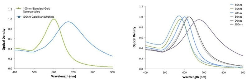
Figure 5. Left - UV-VIS spectra of 100 nm Gold NanoUrchins (blue) and 100 nm standard gold nanoparticles (green). Note the red-shift in the SPR-peak. Right - UV-VIS spectra of Gold NanoUrchins ranging in size from 50 nm to 100 nm in diameter.
Outlook
Gold nanoparticles, owing to their well-established synthetic methodologies, possess well-defined electronic and physical properties, making them highly versatile for a myriad of applications. Furthermore, their surface chemistry is readily modifiable, adding to their appeal. These attributes have propelled gold nanoparticles to the forefront of nanomaterials used extensively in academic research and as a crucial element in point-of-care medical devices and industrial products across the globe. Our extensive portfolio of gold nanoparticles, accessible to researchers worldwide, aims to further enhance their adoption in cutting-edge technological applications.
References
1. Huang D, Liao F, Molesa S, Redinger D, Subramanian V. 2003. Plastic-Compatible Low Resistance Printable Gold Nanoparticle Conductors for Flexible Electronics. J. Electrochem. Soc.. 150(7): G412. https://doi.org/10.1149/1.1582466
2. Stuchinskaya T, Moreno M, Cook MJ, Edwards DR, Russell DA. 2011. Targeted photodynamic therapy of breast cancer cells using antibody-phthalocyanine-gold nanoparticle conjugates. Photochem. Photobiol. Sci.. 10(5):822. https://doi.org/10.1039/c1pp05014a
3. Brown SD, Nativo P, Smith J, Stirling D, Edwards PR, Venugopal B, Flint DJ, Plumb JA, Graham D, Wheate NJ. 2010. Gold Nanoparticles for the Improved Anticancer Drug Delivery of the Active Component of Oxaliplatin. J. Am. Chem. Soc.. 132(13):4678-4684. https://doi.org/10.1021/ja908117a
4. Ali ME, Mustafa S, Hashim U, Che Man YB, Foo KL. 2012. Nanobioprobe for the Determination of Pork Adulteration in Burger Formulations. Journal of Nanomaterials. 20121-7. https://doi.org/10.1155/2012/832387
5. Perrault SD, Chan WCW. 2010. In vivo assembly of nanoparticle components to improve targeted cancer imaging. Proceedings of the National Academy of Sciences. 107(25):11194-11199. https://doi.org/10.1073/pnas.1001367107
6. Peng G, Tisch U, Adams O, Hakim M, Shehada N, Broza YY, Billan S, Abdah-Bortnyak R, Kuten A, Haick H. 2009. Diagnosing lung cancer in exhaled breath using gold nanoparticles. Nature Nanotech. 4(10):669-673. https://doi.org/10.1038/nnano.2009.235
7. Thompson DT. 2007. Using gold nanoparticles for catalysis. Nano Today. 2(4):40-43. https://doi.org/10.1016/s1748-0132(07)70116-0
Aladdinsci: https://www.aladdinsci.com/