Preparation and precautions for mouse lymph node single-cell suspensions
Procedure for preparation of mouse lymph node single cell suspension
1. Dislocate the cervical vertebrae of mice, soak them in 75% alcohol for 5 min, remove the mice, and place them on a sterile operating table with their abdomens facing upwards.
2. Use clippers to cut the skin from the sternum along the median line to the submandibular region, and then cut the skin from the submandibular region toward the roots of the right and left ears. The skin was lifted to the right and left with forceps and fixed with a needle to reveal a pair of large submandibular glands above the sternum. At the upper margin of each of the right and left submandibular glands, yellow anterior cervical lymph nodes were attached. The sternocleidomastoid muscle and the muscle belly were cut and both of their severed ends were lifted to reveal a small deep cervical lymph node to the left and a small deep cervical lymph node to the left and right of the dorsal depth of the left submandibular gland. The lymph nodes are carefully removed with forceps and small ophthalmic scissors.
3. The lymph nodes were removed and immersed in clean PBS solution.
4. Aspirate the culture solution with a sterile 2.5 mL syringe, hold the lymph node with forceps in the left hand and the syringe in the right hand, and carefully insert it into the lymph node and blow it out until the lymph node cells are completely blown out and only white connective and adipose tissue is observed to remain.
5. The blown lymph node cells were filtered through a 200-mesh sieve, collected in a 15 mL centrifuge tube, centrifuged at 300 g for 5 min, and the supernatant was discarded.
6. Lymph node cells were resuspended with cell staining buffer, counted, and the cell concentration was adjusted to 1 × 10⁷/mL.
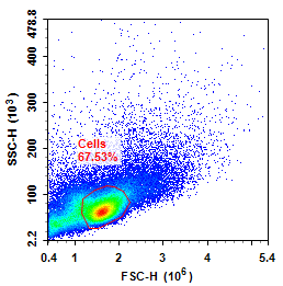
FSC/SSC plot of mouse lymph node cells
Precautions:
1. Cervical lymph nodes
The superficial cervical lymph nodes are located superficial and lateral to the right and left submandibular glands, often surrounded by adipose tissue, and are ovoid in shape. The deep cervical lymph nodes are located deep in the ventral aspect of the right and left sternomastoid and clavicular mastoid muscles, and in the lateral aspect of the muscles below the hyoid bone, i.e., deep in the dorsal aspect of the submandibular abdomen, and they are slightly compressed and bead-shaped. Removal, such as acute experiments can be preceded by puncture of the femoral artery, carotid artery cut off bloodletting, to avoid bleeding during surgery difficult to observe the location of lymph nodes.
2. Axillary lymph nodes
Located in the right and left axillary adipose tissue. It is pear-shaped, cut the chest muscle along the sternum, lift it to the outside and above, peel off the axillary adipose tissue, you can find this pair of lymph nodes with larger size.
3. Arm lymph nodes
The lymph nodes of the arm are located in the superficial subcutaneous connective tissue, close to the right and left side of the biceps muscle belly, in the shape of an oval, and can be found along the subscapularis muscle from the axilla outward.
4. Intrathoracic lymph nodes
The lymph nodes in this area are subcircular, and their number and location vary considerably. They can generally be observed in the following places: in the adipose tissue dorsal to the lateral aspect of the thymus, and posterior to the trachea and the branches of the trachea. To remove them, the sternum is cut with scissors, and the thoracic cavity is opened from side to side to expose the right and left thymus glands located above the heart. Three or four thymic lymph nodes are attached to the medial side of the right and left thymus lobes, opposite the bronchial branches. Because of the similarity in color to the white thymus, care is needed when removing them.
5. Inguinal lymph nodes
Nearly kidney-shaped, they are shallowly located in a trap deep to the left and right gluteal muscles, the head of which is the emanation of the sciatic nerve. The pair of lymph nodes can be found by clipping the anterior-most lumbar and dorsal muscles, peeling back their superficial layers, and gradually lifting them caudally. Because of the small size of this pair of lymph nodes, it is not easy to rediscover them if their local relationship is inadvertently disturbed.
6. Pancreatic lymph nodes
The lymph nodes of the pancreas are a pair of close-packed, nearly spherical lymph nodes with an abdominal blood vessel in the center, located between the medial margins of the right and left kidneys and the abdominal aorta, near the caudal end of the adrenal gland. The hilar lymph nodes are obtained by turning the kidneys medially and peeling away the fatty tissue without damaging the large intra-abdominal vessels. The left lymph node is attached to the ventral side of the renal artery and renal vein and can be easily removed, while the right lymph node is located on the dorsal side of the renal artery and renal vein and cannot be seen or removed. Therefore, if the right kidney is turned towards the left kidney, the right lymph node can be seen attached to the abdominal aorta and can be easily removed.
7. Mesenteric lymph nodes
This is the largest and easiest to find lymph node in all parts of the body of the mouse, which is shaped like an earthworm and extends in the fatty tissue of the mesentery. The cecum is searched for first, and the lymph nodes are removed by cutting the mesenteric tissue along the mesentery upwards.
8. Lumbar lymph nodes
The lumbar lymph nodes are located on either side of the abdominal aorta, above its bifurcation. Both lymph nodes are ovoid in shape, with the left lumbar lymph node located closer to the end.
9. Caudal lymph nodes
The lymph nodes are spherical in shape, situated in the branches of the abdominal aorta, favoring the right side, and are relatively small in size, so it is important to look for them carefully.
10. Stomach lymph nodes
The location of the lymph nodes in the stomach can vary somewhat. Most of them can be found in the lesser curvature as a very small lymph node, and occasionally two lymph nodes can be found in the cardia or the lower esophagus.
11. Axillary lymph nodes
The lymph nodes in the axilla are subglobular, located in the superficial subcutaneous adipose tissue of the right and left axilla, and are not difficult to find. However, when dissecting, the skin should not be separated from the local area, otherwise the anatomical relationship will be disturbed and it is not easy to find.
Number and length of lymph nodes in mice
1. Number of lymph nodes
Only one lymph node is seen in most sites, but two to three lymph nodes, or as many as four, can be seen in the superficial cervical, intrathoracic, gastric, pancreatic, and mesenteric regions, with more variation in the number of intrathoracic lymph nodes. The probability of dissecting lymph nodes varies from site to site. For example, it is difficult to find lymph nodes in the deep cervical region, caudal region or sciatic region, which requires more skillful dissection and careful operation; while it is easier to find lymph nodes in the superficial cervical region, axillary region or mesenteric region.
2. Length of lymph nodes
Most of the lymph nodes have a diameter of 2-3.5 mm, the largest being the mesenteric lymph nodes with a diameter of more than 10 mm, and the smallest being the deep cervical, sciatic and caudal lymph nodes with a diameter of equal to or less than 1 mm; in general, the diameter of normal lymph nodes does not exceed 4 mm.
For more information, visit our website: www.aladdinsci.com.
