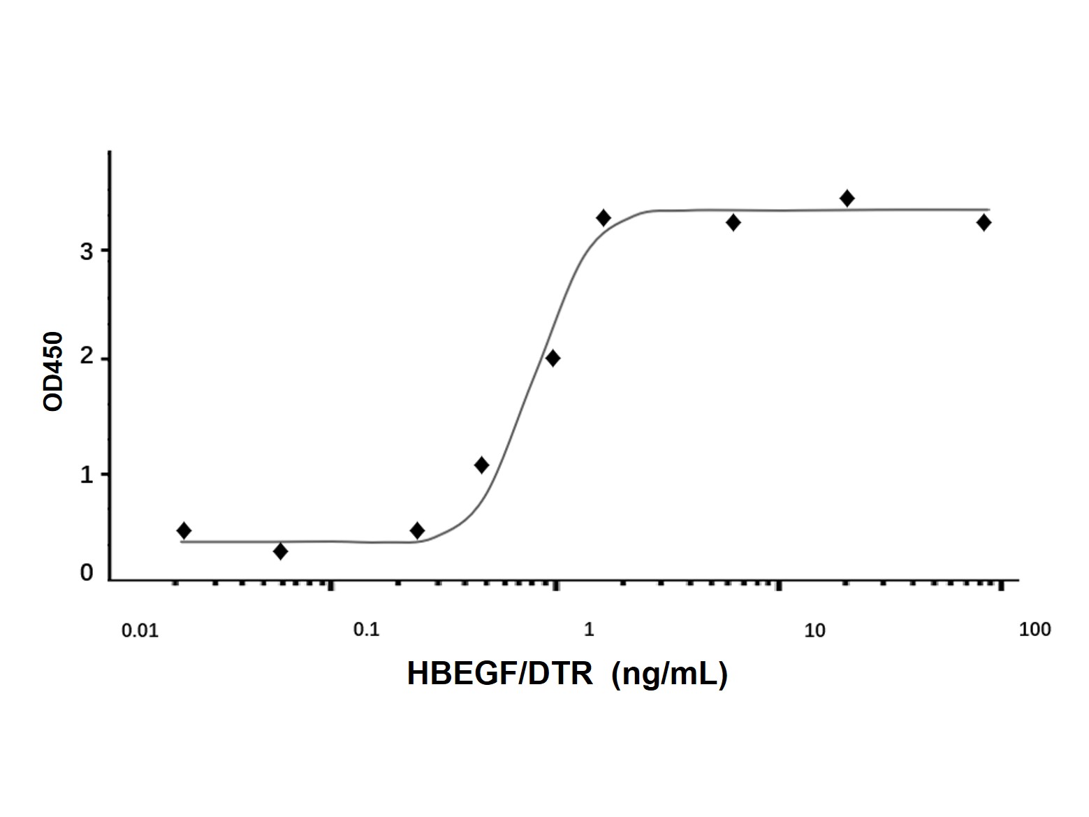
| Product Description | Purity
>97% by SDS-PAGE and HPLC analyses.
Function
May be involved in macrophage-mediated cellular proliferation. It is mitogenic for fibroblasts and smooth muscle but not endothelial cells. It is able to bind EGF receptors with higher affinity than EGF itself and is a far more potent mitogen for smooth muscle cells than EGF. Also acts as a diphtheria toxin receptor.
Background:
Human HB-EGF (Heparin-Binding EGF-like growth factor) is a 12-16 kDa member of the EGF family of peptide growth factors (1-3). Also known as the DTR (diphtheria toxin receptor), it is further classified as a group 2 ErbB ligand based on its ability to activate both the EGF/ErbB1 and ErbB4 receptors (4, 5). HB-EGF is synthesized as a 208 amino acid (aa) type I transmembrane preproprecursor (1, 6). It contains a 19 aa signal sequence, a 43 aa prosegment, an 86 aa mature region (aa 63-148), an 11 aa juxtamembrane cleavage peptide, a 24 aa transmembrane segment, and a 25 aa cytoplasmic tail (aa 184-208). As an integral membrane protein, HB-EGF is expressed as a 19-27 kDa protein in mammalian cells (7-9). The variability in molecular weight (MW) is attributed to heterogeneity in glycosylation and/or the utilization of multiple proteolytic cleavage sites during maturation. Mature HB-EGF is a soluble peptide that arises from proteolytic processing of the transmembrane form. It possesses an EGF-like domain between aa 104-144, and a heparin-binding motif between aa 93‑113. Although the aa range for "mature" HB-EGF is typically stated to be Asp63-Leu148, potential N-terminal start (cleavage) sites also exist at Gly32, Arg73, Val74, Ser77 and Ala82 (8, 10-12). Thus, differential processing (in part) likely accounts for the 16-23 kDa range in MW noted for mammalian-derived mature HB-EGF. Proteases suggested to contribute to HB-EGF processing include TACE, MMP-3 and -7, ADAM-17 and ADAM-12 (11, 13-16). When expressed recombinantly in E.coli, HB-EGF (aa 73-148) runs at 14 kDa in SDS-PAGE; when expressed in Baculovirus, HB-EGF (aa 63-148, 77-148 and 32-148) runs at 18 kDa, 15 kDa, and 19 kDa respectively (8, 12, 17). Over aa 63-148, human HB-EGF- shares 76% and 73% aa sequence identity with rat and mouse HB-EGF, respectively (1, 18). Cells known to express HB-EGF include bronchial epithelium (19), visceral and vascular smooth muscle (20, 21), CD4+ T cells (22), cardiac muscle (23), glomerular podocytes (24), keratinocytes (13) and IL-10-secreting regulatory macrophages (25). As noted earlier, HB-EGF is known to bind to both 170 kDa EGFR and 180 kDa ErbB4, and through heterodimerization, ErbB2 (13, 26). Activity associated with ErbB4 binding appears to be limited to non-mitogenic actions, while EGFR binding induces both mitogenic and non-mitogenic activity.
|
|---|


